Abdominal Anatomy : Abdominal aorta - Wikipedia. Abdominal anatomy seen on ct. Name the planes used for dividing abdominal cavity into regions. Sciency root words make anatomical parts harder to memorize. Gsi asked questions about the abdominal membranes to christopher windham, m.d. Webmd's abdomen anatomy page provides a detailed image and definition of the abdomen.
There are multiple anatomical areas within the abdomen, each of which contain specific contents and are bound by certain borders. In anatomy and physiology, you'll learn how to divide the abdomen into nine different regions and four different quadrants. The abdomen is comprised primarily of the digestive tract and other accessory organs which assist in digestion, the urinary system, spleen, and the abdominal. Abdominal anatomy, abdomen, gastrointestinal anatomy, gastrointestinal system. If you plan to enter a healthcare profession such as nursing, this is something you'll use on the job when performing abdominal assessments (and while documenting).
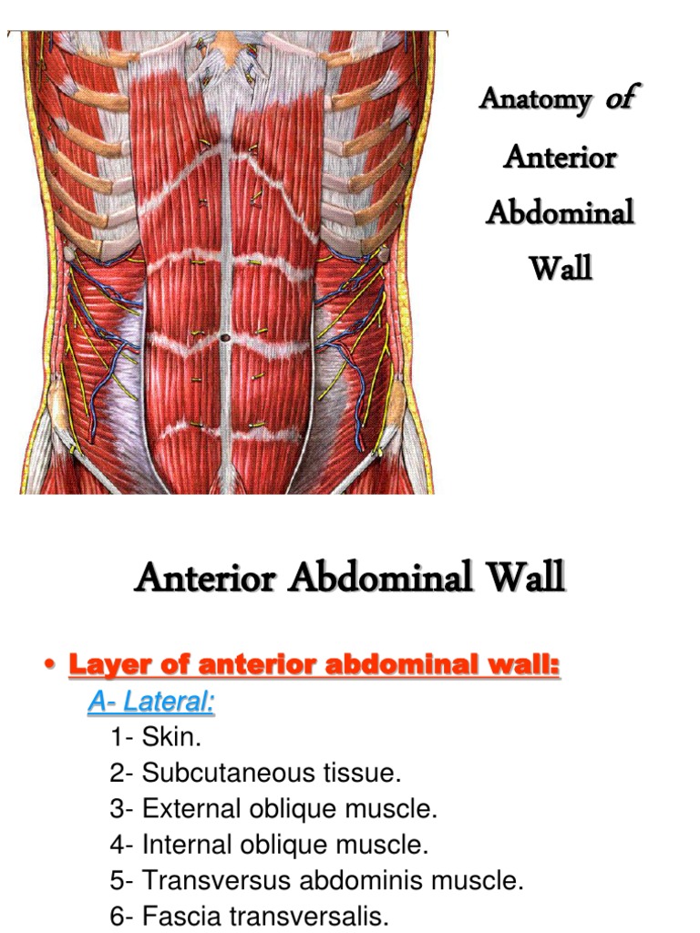
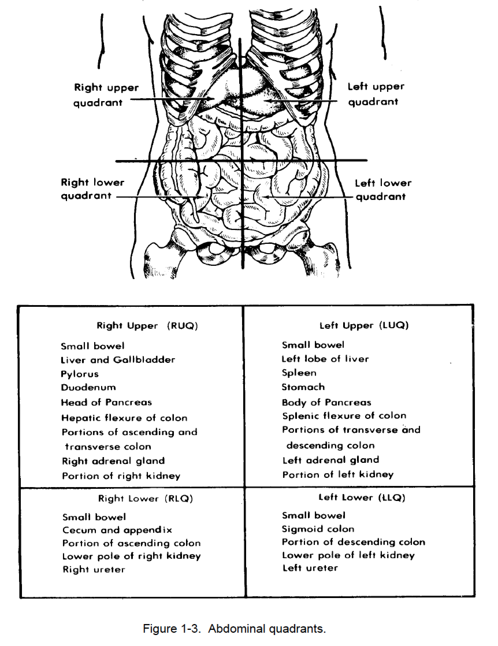
Anterior Abdominal Wall | Abdomen | Human Anatomy from imgv2-2-f.scribdassets.com Divided into 9 regions by two vertical and two horizontal imaginary planes. Inflammation of the covering of the abdominal structures, causing abdominal wall rigidity and severe pain. In anatomy and physiology, you'll learn how to divide the abdomen into nine different regions and four different quadrants. Lee moffitt cancer center & research institute in. The abdominal divisions should be used in conjunction with other diagnostic approaches in order to accurately diagnose a patient's condition. Windham was previously a surgical oncologist in the sarcoma program of the h. The above lines intersect and divide the abdomen into nine regions (clockwise from the top) Radiology basics of abdominal ct anatomy with annotated coronal images and scrollable axial images to help medical students and junior doctors learning anatomy.
Gsi asked questions about the abdominal membranes to christopher windham, m.d.
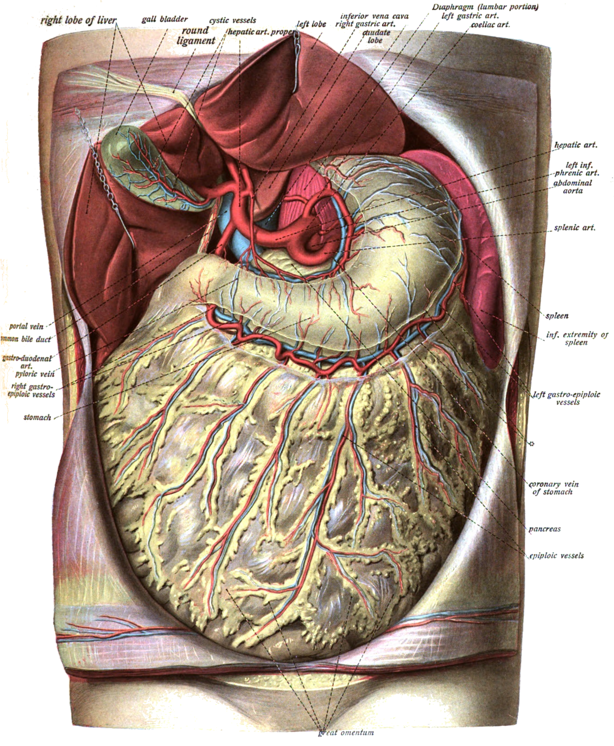
Abdomen, in human anatomy, the body cavity lying between the chest or thorax above and the pelvis below and from the spine in the back to the wall the abdominal organs are supported and protected by the bones of the pelvis and ribcage and are covered by the greater omentum, a fold of peritoneum. Become familiar with the anatomical divisions by exploring the world's most advanced 3d anatomy platform in complete anatomy. Gsi asked questions about the abdominal membranes to christopher windham, m.d. Abdominal anatomy seen on ct. The ovarian vessels enter the. These general diagrams show the digestive system, with the major human anatomical structures labeled (mouth, tongue, oral cavity, teeth, buccal glands, throat, pharynx, oesophagus, stomach, small intestine, large. In order to find the right training and to perform the exercises properly, it is important to know what are the abdominal muscles. Name the planes used for dividing abdominal cavity into regions. Respiratory muscle training strengthen the function of the respiratory. This is a laparoscopic tour of abdominal cavity anatomy. Respiratory muscle training online course: Related online courses on physioplus. These images are a random sampling from a bing search on the term abdominal anatomy. click on the image (or right click) to open the source website in a new browser window.
The abdomen is comprised primarily of the digestive tract and other accessory organs which assist in digestion, the urinary system, spleen, and the abdominal. Two layers in abdomenfatty superficial layer (camper's fascia)deeper membranous layer (scarper's fascia). The ovarian vessels enter the. The abdominal cavity is an ovoid space bounded cephalad by the diaphragm and inferior thoracic margin, caudally by the pelvic brim, posteriorly by the lumbar spine along with quadratus lumborum, psoas major and iliacus, and. The abdomen (colloquially called the belly, tummy, midriff or stomach) is the part of the body between the thorax (chest) and pelvis, in humans and in other vertebrates.
1-16. EXAMINATION OF THE ABDOMEN | Nursing Care Related to the Gastrointestinal and ... from brooksidepress.org Divided into 9 regions by two vertical and two horizontal imaginary planes. The abdomen is comprised primarily of the digestive tract and other accessory organs which assist in digestion, the urinary system, spleen, and the abdominal. Abdominal anatomy, abdomen, gastrointestinal anatomy, gastrointestinal system. Related online courses on physioplus. Learn about its function, parts, abdominal conditions, and abdomen conditions. The ovarian vessels enter the. But with the use of smart technology, you can learn faster and master abdomen anatomy in no time! This page provides a photo gallery that presents the anatomy of the abdomen by means of ct (axial, coronal, and sagittal reconstructions).
Abdominal surface anatomy can be described when viewed from in front of the abdomen in 2 ways:
Sciency root words make anatomical parts harder to memorize. If you plan to enter a healthcare profession such as nursing, this is something you'll use on the job when performing abdominal assessments (and while documenting). Learn about its function, parts, abdominal conditions, and abdomen conditions. Connections between the left and the middle colic the gonadal arteries cross the abdominal ureters approximately halfway between the pelvic inlet and the renal pelvis. The vascular anatomy distal to the middle colic artery and near the splenic flexure is variable. The above lines intersect and divide the abdomen into nine regions (clockwise from the top) This page provides a photo gallery that presents the anatomy of the abdomen by means of ct (axial, coronal, and sagittal reconstructions). The abdominal divisions should be used in conjunction with other diagnostic approaches in order to accurately diagnose a patient's condition. Windham was previously a surgical oncologist in the sarcoma program of the h. The abdominal cavity is an ovoid space bounded cephalad by the diaphragm and inferior thoracic margin, caudally by the pelvic brim, posteriorly by the lumbar spine along with quadratus lumborum, psoas major and iliacus, and. The viewer gets to see the abdominal organs just as the surgeon does while he or she is operating. The xiphoid process and costal. The abdomen (colloquially called the belly, tummy, midriff or stomach) is the part of the body between the thorax (chest) and pelvis, in humans and in other vertebrates.
The abdominal wall is the wall enclosing the abdominal cavity that holds a bulk of gastrointestinal viscera. The ovarian vessels enter the. This muscle forms the anterior and lateral abdominal wall. See more ideas about anatomy, massage therapy, muscle anatomy. The vascular anatomy distal to the middle colic artery and near the splenic flexure is variable.
Greater omentum - Wikipedia from upload.wikimedia.org Lee moffitt cancer center & research institute in. Divided into 9 regions by two vertical and two horizontal imaginary planes. Related online courses on physioplus. The abdominal divisions should be used in conjunction with other diagnostic approaches in order to accurately diagnose a patient's condition. Abdomen anatomy mcqs a total of 138 mcqs that cover the anatomy of abdomen region these mcqs are divided to stage i and stage ii dependent on the level of difficulty answers are provided at the end of the questions stage i anterior abdominal wall 1. Two layers in abdomenfatty superficial layer (camper's fascia)deeper membranous layer (scarper's fascia). In order to find the right training and to perform the exercises properly, it is important to know what are the abdominal muscles. Anatomy of the abdominal wall.
Anatomy of the abdominal wall.
Abdominal surface anatomy can be described when viewed from in front of the abdomen in 2 ways: If you plan to enter a healthcare profession such as nursing, this is something you'll use on the job when performing abdominal assessments (and while documenting). Anatomy of the abdominal wall. Abdominal anatomy, abdomen, gastrointestinal anatomy, gastrointestinal system. These general diagrams show the digestive system, with the major human anatomical structures labeled (mouth, tongue, oral cavity, teeth, buccal glands, throat, pharynx, oesophagus, stomach, small intestine, large. Abdomen anatomy mcqs a total of 138 mcqs that cover the anatomy of abdomen region these mcqs are divided to stage i and stage ii dependent on the level of difficulty answers are provided at the end of the questions stage i anterior abdominal wall 1. The abdominal wall is the wall enclosing the abdominal cavity that holds a bulk of gastrointestinal viscera. Abdominal anatomy seen on ct. The above lines intersect and divide the abdomen into nine regions (clockwise from the top) This page provides a photo gallery that presents the anatomy of the abdomen by means of ct (axial, coronal, and sagittal reconstructions). The vascular anatomy distal to the middle colic artery and near the splenic flexure is variable. Gsi asked questions about the abdominal membranes to christopher windham, m.d. Two layers in abdomenfatty superficial layer (camper's fascia)deeper membranous layer (scarper's fascia).
Share :
Post a Comment
for "Abdominal Anatomy : Abdominal aorta - Wikipedia"

Post a Comment for "Abdominal Anatomy : Abdominal aorta - Wikipedia"